Consent for Cookie Use We use cookies to improve your experience on our websites. You can learn more about what information we collect and how we use it on our Internet Privacy Statement.
Positron emission tomography (PET), Opens dialog is a test that uses a special type of camera and a tracer, Opens dialog (radioactive substance) to look at organs in the body. The tracer usually is a special form of a substance (such as glucose) that collects in cells that are using a lot of energy, such as cancer cells.
During the test, the tracer liquid is put into a vein (intravenous, or IV, Opens dialog) in your arm. The tracer moves through your body, where much of it collects in the specific organ or tissue. The tracer gives off tiny positively charged particles (positrons). The camera records the positrons and turns the recording into pictures on a computer.
Evaluate the extent of some cancers, especially lymphoma, Opens dialog or cancers of the head and neck, brain, lung, colon, or prostate. In its early stages, cancer may show up more clearly on a PET scan than on a CT scan or an MRI.
Determine whether a growth in an organ or in tissue is likely to be cancer, such as a growth in lung tissue.
See how advanced a cancer is and whether it has spread to another area of the body (metastasized). It is often necessary to do both CT and PET scans to evaluate cancer.
Help a doctor choose the best treatment for cancer or to see how well treatment is working. PET scans may also be done to see whether surgery can be done to remove a tumor.
Help diagnose Alzheimer's disease, Opens dialog when the symptoms are not clear or when a person has dementia symptoms at a young age (usually younger than 65).footnote 1 This is called amyloid imaging.
You have diabetes. If you take medicine to control diabetes, you may need to take less than your normal dose. Talk with your doctor about how much medicine you should take.
You take any medicines, supplements, or herbal remedies. You may need to stop taking some medicines or change your dose before this test.
You are or might be pregnant.
You are breastfeeding. The radioactive tracer used in this test can get into your breast milk. Do not breastfeed your baby for 2 days after this test. During this time, you can give your baby breast milk you stored before the test, or you can give formula. Discard the breast milk you pump for 2 days after the test.
You have a fear of enclosed spaces.
Do not smoke or drink caffeine or alcohol for 24 hours before this test.
Do not eat or drink (except water) for at least 6 hours before this test.
You may be asked to sign a consent form.
Talk to your doctor about any concerns you have regarding the need for the test, its risks, how it will be done or what the results mean. To help you understand the importance of this test, fill out the medical test information form .
Information about Positron Emission Tomography (PET)
Positron Emission Tomography (PET)—HowItIsDone
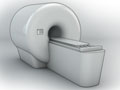
A positron emission tomography (PET) scan is done in a hospital nuclear medicine department or at a special PET center by a radiologist, Opens dialog or nuclear medicine specialist, Opens dialog and a technologist. You will lie on a table that is hooked to a large scanner, camera, and computer.
During the test
The radioactive tracer is usually given in a vein (IV). You may need to wait 30 to 60 minutes for the tracer to move through your body. During this time, you may need to avoid moving and talking.
The PET scanner, which is shaped like a doughnut, moves around you. The scanned pictures are sent to a computer screen so your doctor can see them. Many scans are done to make a series of pictures. It is very important to lie still while each scan is being done. At some medical centers, a CT scan will be done at the same time.
For a PET scan of the brain, you will lie on a bed. You may be asked to read, name letters, or tell a story, depending on whether speech, reasoning, or memory is being tested. During the scan, you may be given earplugs and a blindfold (if you do not need to read during the test) to wear for your comfort.
During the test, you will be alone in the scanner room. The technologist will watch you through a window and you will be able talk to him or her through a two-way intercom at all times.
The test takes 1 to 3 hours.
After the test
After the test, drink lots of fluids for the next 24 hours to help flush the tracer out of your body.
Positron Emission Tomography (PET)—HowItFeels
You will not feel pain during the test. The table you lie on may be hard and the room may be cool. It may be difficult to lie still during the test.
You may feel a quick sting or pinch when the IV is put in your arm. The tracer is unlikely to cause any side effects. If you don't feel well during or after the test, tell the person who is doing the test.
In rare cases, some soreness or swelling may develop at the IV site where the radioactive tracer was put in. Apply a moist, warm compress to your arm.
Anytime you're exposed to radiation, there's a small chance of damage to cells or tissue. That's the case even with the low-level radioactive tracer used for this test. But the chance of damage is very low compared with the benefits of the test.
Most of the tracer will leave your body through your urine or stool within a day. So be sure to flush the toilet right after you use it, and wash your hands well with soap and water. The amount of radiation in the tracer is very small. This means it isn't a risk for people to be around you after the test.
Positron Emission Tomography (PET)—Results
Positron emission tomography (PET) is a test that uses a special type of camera and a tracer, Opens dialog (radioactive substance) to look at organs in the body.
The radiologist, Opens dialog may discuss preliminary results of the PET scan with you right after the test. Complete results are usually available in 1 to 2 days.
Positron emission tomography (PET)
Normal:
Blood flow is normal and organs are working well. The flow and pattern of the tracer shows normal distribution in the body.
Having recently had surgery, a biopsy, chemotherapy, or radiation therapy.
Citations
Johnson KA, et al. (2013). Appropriate use criteria for amyloid PET: A report of the Amyloid Imaging Task Force, the Society of Nuclear Medicine and Molecular Imaging, and the Alzheimer's Association. Alzheimer's and Dementia, 9(1): e1–e16.
Current as of: July 31, 2024
Author: Ignite Healthwise, LLC Staff Clinical Review Board All Healthwise education is reviewed by a team that includes physicians, nurses, advanced practitioners, registered dieticians, and other healthcare professionals.
Clinical Review Board All Healthwise education is reviewed by a team that includes physicians, nurses, advanced practitioners, registered dieticians, and other healthcare professionals.
Johnson KA, et al. (2013). Appropriate use criteria for amyloid PET: A report of the Amyloid Imaging Task Force, the Society of Nuclear Medicine and Molecular Imaging, and the Alzheimer's Association. Alzheimer's and Dementia, 9(1): e1–e16.
This information does not replace the advice of a doctor. Ignite Healthwise, LLC, disclaims any warranty or liability for your use of this information. Your use of this information means that you agree to the Terms of Use. Learn how we develop our content.