Computed Tomography (CT) Scan of the Head and Face
Test Overview
A computed tomography (CT) scan uses X-rays to make pictures of the head and face.
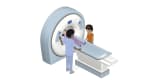
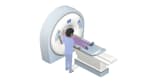
During the test, you will lie on a table that is attached to the CT scanner, which is a large doughnut-shaped machine. Your head will be positioned inside the scanner. The CT scanner sends X-rays through the head. Each rotation of the scanner provides a picture of a thin slice of the head and face. One part of the scanning machine can tilt to take pictures from different positions. All of the pictures are saved as a group on a computer. They also can be printed.
In some cases, a dye called contrast material may be put in a vein (I.V.) in your arm. The dye makes structures and organs easier to see on the CT pictures. The dye may be used to check blood flow and look for tumors, areas of inflammation, Opens dialog, or nerve damage.
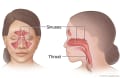
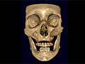
A CT scan of the head can give some information about the eyes, facial bones, air-filled cavities (sinuses) within the bones around the nose, and the inner ear. If these areas are of concern, a specific CT scan of the area is usually done.
A CT scan of the head may be used to evaluate headaches.
Find the cause of symptoms, such as confusion, paralysis, numbness, vision problems, vertigo, or headaches, that might mean a brain injury, a brain tumor, a ruptured aneurysm, or bleeding inside the head.
Look for problems of the middle ear bones and the auditory nerve.
Help plan for surgery.
Find damage caused by a stroke and to help find the best treatment for the cause of a stroke.
Find the cause of a loss of consciousness or a changing level of consciousness.
Check on the success of treatment or surgery for a brain tumor.
Plan for surgery to rebuild parts of the face that were damaged.
In general, there's nothing you have to do before this test, unless your doctor tells you to.
Tell your doctor if you get nervous in tight spaces. You may get a medicine to help you relax. If you think you'll get this medicine, be sure you have someone to take you home.
Information about Computed Tomography (CT) Scan of the Head and Face
You may need to take off any jewelry, glasses, and hearing aids. Wear comfortable, loose-fitting clothes.
During the test, you will lie on a table that is attached to the CT scanner. Straps will hold your head still, but your face will not be covered.
The table slides into the round opening of the scanner, and the scanner moves around your body. The table will move while the scanner takes pictures. You may hear a click or buzz as the table and scanner move. It is very important to lie still during the test.
You may be alone in the scan room. But a technologist will watch you through a window and talk with you during the test.
How long the test takes
The test will take about 30 to 60 minutes. Most of this time is spent getting ready for the scan. The actual test only takes a few minutes.
Watch
The test will not cause pain, but some people feel nervous inside the CT scanner.
If a medicine to help you relax (sedative) or dye is given through an I.V., you may feel a quick sting or pinch when the I.V. is started. The dye may make you feel warm and flushed and give you a metallic taste in your mouth. Some people feel sick to their stomach or get a headache. Tell the technologist or your doctor how you are feeling.
The chance of a CT scan causing a problem is small.
If you breastfeed and are concerned about whether the contrast material used in this test is safe, talk to your doctor. Most experts believe that very little dye passes into breast milk and even less is passed on to the baby. But if you are concerned, you can stop breastfeeding for up to 24 hours after the test. During this time, you can give your baby breast milk that you stored before the test. Don't use the breast milk you pump in the 24 hours after the test. Throw it out.
There is a risk of damage to cells or tissue from being exposed to radiation, including the small amounts used in CTs, X-rays, and other medical tests. Over time, exposure to radiation may cause cancer and other health problems. But in most cases, the risk of getting cancer from being exposed to small amounts of radiation is low. It is not a reason to avoid these tests for most people.
Complete results usually are ready for your doctor in 1 to 2 days.
CT scan of the head and face
Normal:
The brain and blood vessels and bones of the skull and face are normal in size, shape, and position.
There are no foreign objects or growths.
No bleeding or collections of fluid are found.
Abnormal:
A growth, such as a tumor, or bleeding is found in or around the brain. Foreign objects, such as glass or metal fragments, are found. The bones of the skull or face are broken (fractured) or look abnormal. Nerves leading to or from the brain are damaged or pinched.
A collection of fluid is found, which may mean bleeding in or around the brain.
An aneurysm is found.
The openings in the brain (ventricles) through which cerebrospinal fluid flows into the spine are enlarged. An area of the brain shows swelling (edema) or other changes that may mean a stroke.
Author: Ignite Healthwise, LLC Staff Clinical Review Board All Healthwise education is reviewed by a team that includes physicians, nurses, advanced practitioners, registered dieticians, and other healthcare professionals.
This information does not replace the advice of a doctor. Ignite Healthwise, LLC, disclaims any warranty or liability for your use of this information. Your use of this information means that you agree to the Terms of Use. Learn how we develop our content.