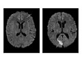
Magnetic resonance imaging (MRI) is a test that uses a magnetic field and pulses of radio wave energy to take pictures of the head. In many cases, MRI gives information that can't be seen on an X-ray, ultrasound, or computed tomography (CT) scan.
For an MRI of the head, you lie with your head inside a special machine (scanner) that has a strong magnet. The MRI can show tissue damage or disease, such as infection or inflammation, or a tumor, stroke, Opens dialog, or seizure, Opens dialog. Information from an MRI can be saved and stored on a computer for more study. Photographs or films of certain views can also be made.
In some cases, a dye (contrast material) may be used during the MRI to show pictures of structures more clearly. The dye may help show blood flow, look for some types of tumors, and show areas of inflammation.
An MRI of the head may be used to look for the cause of headaches.
Magnetic resonance imaging (MRI) of the head is done to:
Look for the cause of headaches.
Help diagnose a stroke or blood vessel problems in the head. Problems with blood vessels may include an aneurysm or abnormal twisted blood vessels that are present at birth (this is called an arteriovenous [AV] malformation).
Check blood flow or blood clots to the brain. MRI can show bleeding in or around the brain.
Check symptoms of a known or suspected head injury.
Look for tumors, infections, an abscess, or conditions of the brain or brain stem, such as encephalitis or meningitis.
Check the eyes, the nerves from the eyes to the brain (optic nerves), the ears, and the nerves from the ears to the brain (auditory nerves).
Look for problems of the pituitary gland.
Investigate or follow a finding seen on another test.
In general, there's nothing you have to do before this test, unless your doctor tells you to.
Tell your doctor if you get nervous in tight spaces. You may get a medicine to help you relax. If you think you'll get this medicine, be sure you have someone to take you home.
Information about Magnetic Resonance Imaging (MRI) of the Head
You may have contrast material (dye) put into your arm through a tube called an I.V.
You will lie on a table that's part of the MRI scanner.
The table will slide into the space that contains the magnet.
Inside the scanner, you will hear a fan and feel air moving. You may hear tapping, thumping, or snapping noises. You may be given earplugs or headphones to reduce the noise.
You will be asked to hold still during the scan. You may be asked to hold your breath for short periods.
You may be alone in the scanning room. But a technologist will watch through a window and talk with you during the test.
How long the test takes
The test usually takes 30 to 60 minutes but can take as long as 2 hours.
You won't have pain from the magnetic field or radio waves used for the MRI test. But you may be tired or sore from lying in one position for a long time.
If a contrast material is used, you may feel some coolness when it is put into your I.V.
In rare cases, you may feel:
Tingling in the mouth if you have metal dental fillings.
Warmth in the area being checked. This is normal. Tell the technologist if you have nausea, vomiting, a headache, dizziness, pain, burning, or breathing problems.
There are no known harmful effects from the strong magnetic field used for an MRI. But the magnet is very powerful. It may affect any metal implants or other medical devices you have.
Risks from contrast material
Contrast material that contains gadolinium may be used in this test. But for most people, the benefit of its use in this test outweighs the risk. Be sure to tell your doctor if you have kidney problems or are pregnant.
There is a slight chance of an allergic reaction if contrast material is used during the test. But most reactions are mild and can be treated using medicine.
If you breastfeed and are concerned about whether the contrast material used in this test is safe, talk to your doctor. Most experts believe that very little dye passes into breast milk and even less is passed on to the baby. But if you are concerned, you can stop breastfeeding for up to 24 hours after the test. During this time, you can give your baby breast milk that you stored before the test. Don't use the breast milk you pump in the 24 hours after the test. Throw it out.
The radiologist may tell you some of the results of the MRI right after the test. Full results are sent to your doctor or specialist in 1 to 2 days.
Normal
All structures of the head—the brain, its vessels, spaces, nerves, and surrounding structures—are normal.
No abnormal growths, such as tumors, in or around the brain are present.
No bleeding, abnormal blood vessels (AV malformations), abnormal pockets of fluid, blockage in the flow of blood, or bulges in the blood vessels (aneurysm) are present.
No signs of infection or inflammatory disease, such as encephalitis or meningitis, are present.
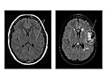
Abnormal
Tumors in the brain or in areas outside the brain, such as an acoustic neuroma, are present.
Bleeding or swelling (edema) in or around the brain is present.
Areas of infection or inflammatory disease, such as encephalitis or meningitis, are present.
Abnormal areas in the brain may mean that certain diseases, such as Huntington's disease, Opens dialog, multiple sclerosis, or Parkinson's disease, are present.
Bulges or weak areas (aneurysms) or abnormal blood vessels (such as an AV malformation) are present.
Author: Ignite Healthwise, LLC Staff Clinical Review Board All Healthwise education is reviewed by a team that includes physicians, nurses, advanced practitioners, registered dieticians, and other healthcare professionals.
This information does not replace the advice of a doctor. Ignite Healthwise, LLC, disclaims any warranty or liability for your use of this information. Your use of this information means that you agree to the Terms of Use. Learn how we develop our content.